| | | | | the mucosae play a very important role in the immunological defence of pigs, as a large number of pathogens use the mucosae as a means of entry. | | | Mucous associated lymphoid tissue (MALT) is part of the immune system even though it works independently of the systemic system. It is responsible for protecting the pig's the mucosae from attack from pathogens in both primary and secondary responses. It is made of nodes of lymphoid tissue which, according to their location, are called: GALT and BALT
| |
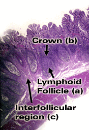
The acronym GALT stands for "Gut Associated Lymphoid Tissues". GALT is made of all the lymphoid tissue found on the walls of the intestines (lymph nodes, Peyer patches, isolated lymph follicles). (Secondary lymphoid organs)
The acronym BALT stands for "Bronchus Associated Lymphoid Tissues". It is made of all lymphoid tissue (tonsils, lymph nodes, lymph follicles) found in the respiratory mucosae from the nasal cavities to the lungs.
A particular feature of the BALT in pigs compared to rodents or humans is the high presence of very active intravascular macrophages in the lungs.
|
|
| IMMUNITY OF THE MUCOSAE.
The large amount of lymphoid tissue in the mucosae of the pig proves the importance of this tissue in defence mechanisms against infections.
This lymphoid tissue is strategically distributed in the following areas:
The initiation or inducting areas of the mucous membrane immune response have similar elements to those of the systemic immune system for uptake of antigens and initiation of the immune response. With the sole exception of the M cells, which are epithelial cells specialized in the transportation of antigens, all other components (presenting-cells, and T and B lymphocytes) act in a similar manner to the systemic system. These cell components are found in the tonsils, Peyer's patches, lymph nodes and diffuse lymphoid tissue. In summary, contact with the antigen, transportation, processing and T and B lymphocyte presentation take place in the GALT and BALT areas (secondary lymphoid organs of the pig) |
|
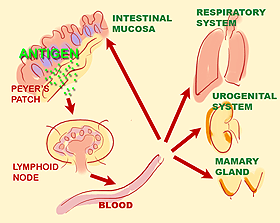
Stimulation of BALT or GALT mucous membrane lymphoid tissue. This mechanism means that even if antigenic stimulation has been local, the immune response is generalized.
|
Antigens usually enter the animal through inhalation or ingestion. Through a process related to M cells, ACP or directly by B lymphocytes (similar to cell cooperation), the B cells (mainly precursors of IgA) and T lymphocytes are stimulated. The cells stimulated in these initiation areas leave the area through the bloodstream, migrating towards the various effector areas.
This mechanism permits a generalized immune response in the animal, even if antigenic stimulation has been at local level. This is known as a generalized secreting immune response. |
|

Diagram of the structure of IgA immunoglobulin |
Most of the immune cells in the effector areas are T lymphocytes, which are located between the epithelial cells (intraepithelial lymphocytes) and underneath them in the lamina propria. These are mainly CD 8 (77%) and CD 4 g-d. B lymphocytes which can react with the antigen are also present. The plasma cells are primarily producers of IgA immunoglobulin isotype, found mainly in the lymph nodes and in the diffuse plasma cells. These are found in the intestinal and respiratory walls. These cells are essential in the mucous membrane immune response, secreting around 80% of the IgA produced, with the sole exception of the tonsils, where the immunoglobulin synthesized in the largest amount is IgG followed by IgA. |
|
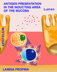
ANTIGENIC PRESENTATION IN THE INDUCTOR AREA OF THE MUCOUS MEMBRANES. Antigens penetrating the enterocytes are quickly destroyed by the lysosomes. However, antigens captured by the M cells are transported without being broken down and are presented to intraepithelial lymphocytes. They are then passed to the lymph nodes.
|
Transportation of antigens to the inductor areas (Peyer's patches and lymphoid follicles) is mainly carried out by M cells. The M cells are epithelial cells specialized in the transportation of antigens. They do not act enzymatically on antigens. The M cells capture antigens in the intestinal lumen and transport them completely intact for presentation to intraepithelial lymphocytes (within the cell itself) or pass through an intercellular space towards the cell fluid and present the antigen to the presenting cells (macrophages, dendritic cells and B lymphocytes) present in the subepithelial space or in the lamina propria. Activation mechanisms at lamina propria level follow a similar pattern to that of cell cooperation described above.
|
|
Diagram of the structure of IgA immunoglobulin |
Most of the immune cells in the effector areas are T lymphocytes, which are located between the epithelial cells (intraepithelial lymphocytes) and underneath them in the lamina propria. These are mainly CD 8 (77%) and CD 4 g-d. B lymphocytes which can react with the antigen are also present.
The plasma cells are primarily producers of IgA immunoglobulin isotype, found mainly in the lymph nodes and in the diffuse plasma cells. These are found in the intestinal and respiratory walls. These cells are essential in the mucous membrane immune response, secreting around 80% of the IgA produced, with the sole exception of the tonsils, where the immunoglobulin synthesized in the largest amount is IgG followed by IgA. |
|
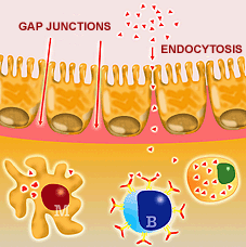
The antigen in the effector area, which it enters by endocytosis or through the tight junctions. |
Antigens presentation can also take place in the effector areas, although the entrance mechanism is usually different to that of the inductor areas. The antigen can enter the effector areas by endocytosis by means of the epithelial cells or by crossing areas known as tight junctions. Capture and presentation is carried out by macrophages, M cells or B lymphocytes, and the subsequent stages follow a procedure as described previously.
The induction capacity of an immune response at mucous membrane level generally requires a greater amount of antigen and at times a greater number of immunizations that that of the systemic system, above all in oral immunizations.
This is due to the many enzymatic changes and degradations suffered by antigens taking this route into the organism. This mechanism is good for the immune defence of the animal, but must be taken into account when designing oral vaccines. In chapter 8 we will see that several strategies exist to induce a good immune response by oral administration. Nevertheless it is generally easier to induce immunity in the respiratory mucosae through oral immunization than inducing an immune response in the intestinal mucosae through nasal immunization.
|
|
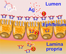
DIAGRAM OF IgA ACTIVITY. IgA can act at intestinal lumen level, preventing attachment of viruses and/or bacteria to the epithelium (1), neutralizing a virus within the enterocytes (2) and finally, in the cell fluid (3).
|
IgA (Chapter 4) plays a vitally important role in the immune response of the mucosae. Its dimeric or tetrameric configuration provides it with 4 to 8 antigen binding sites, making it tremendously effective against different bacterial antigens by means of ADCC type reactions. IgA is not a bactericide, but it does, however, have a great capacity for the neutralization of some viruses, even within epithelial cells. It is the only immunoglobulin capable of acting within a cell, but above all its main activity in the defence of mucosae is preventing bacteria and virus from attaching to the surface of the epithelium. IgA can therefore act in three places and in different ways. Firstly, it can bind the antigen in the intestinal lumen to prevent viruses and/or bacteria from attaching to the surface of the epithelium, secondly, it can act within the enterocytes, and finally, in the cell fluid.
A further example of the importance of this immunoglobulin in porcine defence mechanisms is seen in the fact that in pigs, 85% of the cells that contain immunoglobulins in the lamina propria of the intestine contain IgA. |
| |