- They act as antigen-presenting cells.
- They produce antibodies.
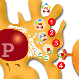
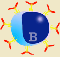
B lymphocytes have SLA II in their membranes, so can therefore recognise the antigen, even in its native form (free antigen without the need to be associated to SLA or having been transformed) by means of its surface immunoglobulins.
| | 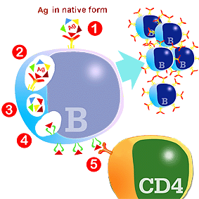
Diagram of Ag presentation by B lymphocytes. Reaction with Ag in its native form (1); internalization of Ag (2); fragmentation of Ag (3); binding to SLA II (4) and expression in the membrane (5)
|
How is B lymphocyte activated?
The antigen in its native form reacts directly with the immunoglobulins in the membrane of the B lymphocytes, without the intervention of presenting cells or T lymphocytes. Next, the antigen is internalized, fragmented, associated to SLA II and expressed in the membrane of the B lymphocyte itself. In summary, it acts in a similar way to the other APCs, with two main differences: a) The antigen can be recognised in its native form and in a specific form by means of surface immunoglobulin and b) There is no phagocytosis.
Once the antigen is expressed in the membrane of the B lymphocytes, if there is appropriate stimulus from a CD4+ lymphocyte, the production of immunoglobulins can start.This collaboration is essential for the initiation of the production of antibodies against most antigens. The collaboration between the B and CD4+ lymphocytes takes place when both cells make contact thanks to SLA II and the antigen, as well as the release of cytokines, above all interleukin 4.
|
A B lymphocyte stimulated by an antigen changes its structure and function. The B lymphocyte divides, producing several clones, and is then transformed, developing rough endoplasmic reticulum, finally becoming a plasma cell.
|
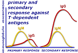
Antibody production starts with IgM and next, if antigenic stimulation continues, the constant region of the mu chains (IgM) is changed to gamma (IgG) and epsilon (IgE) or alpha (IgA), but the original variable regions (those which bind to the antigen) are maintained. (Chapter 4) This process is similar to that originated by antigen presentation by the presenting cells, but it is more efficient as the B lymphocytes only react with the antigen, whilst the macrophages phagocyte all kinds of particles and in addition require a smaller quantity of antigen.
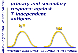
The only exception to this system is seen in T independent antigens which can produce antibodies without the collaboration of T lymphocytes. These antigens, generally present in some bacteria, are usually polysaccharides or lipopolysaccharides, NOT PROTEINS. These can react directly with the BcR by crosslinking, stimulating several BcR at the same time and inducing sufficient stimulus to start antibody induction. However they are only capable of inducing IgM and no other immunoglobulin. |
|
|
|
| Cooperation between APC and T and B lymphocytes. Production of antibodies through the presentation of antigens by phagocytic presenting cells.
The antigens can also be captured by macrophages or dendritic cells which act as antigen-presenting cells. Along with the B lymphocytes, these APC cells belong to an exclusive group of immune system cells expressed in their SLA II membrane (every nucleated cell presents SLA I, but not SLA II). Therefore they are the only ones capable of presenting antigens (epitopes) to the CD 4+ T helper cells.
|
The uptake of these antigens is carried out in this case by a non-specific mechanism (as opposed to B lymphocytes) which usually needs a large amount of antigenic material. The antigen is engulfed within a vesicle (1) and transported to the lysosomes where it is broken down by the various enzymes (2). Fragments of the antigen, once digested, are associated by non-covalent bonds to SLA II (3) and transported to the macrophage membrane (4) where they are recognised by the CD 4+ T lymphocytes (the TcR receptors cannot recognise antigens which are not associated to SLA).
|
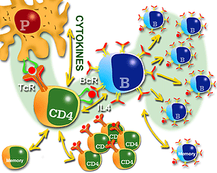
Diagram of cell cooperation for antigen presentation by presenting cells (macrophages or dendritic cells)
|
When these cells interact (APC and T lymphocytes), both are activated and various cytokines and their specific receptors are released. This enables the stimulation and proliferation of CD4+ T lymphocytes (clonal expansion of CD4+). At this time the CD 4+ lymphocytes can stimulate the B lymphocytes to produce antibodies. This stimulation is mediated by SLA II and the antigen (both cells should recognise at least the different epitopes of the same antigen) and is promoted by the release of interleukin 4 (IL-4). The activation of CD4+ T lymphocytes therefore requires the action of antigen-presenting cells (APC) and the type II histocompatibility antigen (SLA II).
The B lymphocytes activated by these mechanisms undergo proliferation. Some of the cells transform into antibody-secreting plasma cells and the remaining B lymphocytes remain as memory cells. The memory cells are long-living lymphocytes, as opposed to conventional lymphocytes and plasma cells which have a very short life. This long life is controlled by a gene known as bcl-2 which is only present in memory lymphocytes. |
|
Characteristics of the primary response:
Characteristics of the secondary response:
|
|
|
When an immune system encounters an antigen for the first time, a type of reaction called a primary response occurs. The primary response mainly takes place in the lymph nodes and in the spleen. During this primary response, memory lymphocytes are produced which remember each epitope's structure in the event of future infections.
When an animal has been in contact with a particular antigen and has produced memory lymphocytes, an immune response called a secondary response will occur the next time the antigen is encountered.
During a secondary response, levels of IgM are similar to those of a primary response, but the IgC levels are much higher and remain longer. During the secondary response other immunoglobulins are produced, such as IgA and IgE. The secondary response mainly takes place in the bone marrow, followed by the spleen and lymph nodes. In T independent antigens the response pattern is the same for both the primary and secondary response, and only the immunoglobulin IgM is produced in both cases. |
|