This group of cells contains:
|
|
| MACROPHAGES |
GRANULOCYTES
- Neutrophils
- Basophils
- Eosinophils
|
| DENDRITIC CELLS. |
|
| | | |
|
Diagram of the structure of a macrophage. |
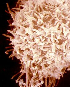
Monocytes-macrophages. These are large cells with a diameter of 15 mm, large cytoplasm and single nucleus. They can be: spherical, kidney-shaped or lobulated. A highly developed Golgi apparatus can be observed in the cytoplasm, as well as rough endoplasmic reticulum, mitochondria and a large number of lysosomes, which are very rich in hydrolytic enzymes, proteases and lipases. These factors indicate their great capacity to synthesize and secrete proteins.
They originate from the myeloid lineage, firstly undergoing transformation into promonocytes in the bone marrow, then into monocytes in the peripheral blood and finally into macrophages in the different organs. |
|
|
ANTIGEN-PRESENTING CELLS: |
| PHAGOCYTIC: |
-
Monocytes.
-
Macrophages.
-
Dendritic cells.
|
| NON-PHAGOCYTIC: |
|
|
|
ROLE OF THE PHAGOCYTIC CELLS.
Two groups of cells can be differentiated within phagocytic cells
1.- Monocytes-macrophages can phagocytize repeatedly and along with dendritic cells and B lymphocytes, make up part of the exclusive group of antigen-presenting cells (APC).
2. The granulocytes: neutrophils, basophils and eosinophils are fast phagocytic cells, but are not able to do so repetitively. They do not present antigens. (They do not express SLA II)
Monocytes-macrophages, alongside dendritic cells and B lymphocytes, are the only porcine cells that express SLA II histocompatibility antigens on their surface and as such are the only cells capable of presenting antigens to the CD4+ helper cells. This cell group is known as Antigen-presenting Cells or APC
Monoclonal antibodies are still used to differentiate and study monocytes-macrophages. At the last symposium on differentiation of cells of the porcine immune system, held in 1998, three monoclonal antibodies were defined for the study of porcine macrophages. These were named Swine Workshop Cluster plus a number. The most commonly used monoclonal antibodies these days are SWC1 and SWC9, which mainly differentiate monocytes and macrophages, and SWC3 which reacts with monocytes, differentiated macrophages and granulocytes.
|
MONOCLONAL ANTIBODIES FOR THE STUDY OF MACROPHAGES AND GRANULOCYTES. |
| SWC1 |
SWC9 |
SWC3 |
|
|
Cells in suspension which have had monoclonal antibodies added pass through a very fine tube in a single line of cells. They are read by the laser and the light beam; the former identifies the labelled antibodies and the latter the size of the cells.
|
Thanks to these markers, it has recently been possible to determine that a monocyte is SCW1+SWC9-. When transformed into a macrophage, however, it becomes SWC1-SWC9+. Studies on peripheral blood monocytes can be performed using flow cytometry, whilst inmunohistochemical techniques are used to study tissues (How are cells studied?).
MACROPHAGES ARE FOUND IN MANY ORGANS. |
| Liver: Kupffer cells |
| Bone tissue: Osteoclasts |
| Nervous tissue: Microglia cells |
| Skin: Langerhans cells |
| Spleen: Dendritic cells. |
| Lymphatic system: Monocytes, macrophages, dendritic cells |
| Lungs: Alveolar macrophages |
| Connective tissue: Histiocytes |
As well as different morphology depending on the organ where they are found, macrophages present different activity according to their level of maturity, activation and location.
The following activities should be emphasised:
| PHAGOCYTIC. (antimicrobial and anti-tumour activity) |
ANTIGEN PRESENTATION. Stimulation of lymphocytes |
SYNTHESIS of a large number of CYTOKINES. |
Macrophages carry out functions of phagocytosis and lysis of microorganisms and infected and/or tumoural cells, either directly (natural or innate immunity) or through Fc receptors for immunoglobulins, or complement receptors. As presenting cells, they participate in the induction of adaptive immunity, capturing and processing antigens in order to present them to T lymphocytes (CD 4), later associated to SLA II. Finally, the macrophages synthesize a large number of cytokines: Interleukines: 1, 2, 6, 12, interferon a and b and tumour necrosis factor-alpha cell (TNF-a), as well as the complement components C2, C3, C4 and C5 and several enzymes.
|
|
| MACROPHAGE RECEPTORS |
|
| Antibody receptor |
| Receptor for several complement components |
| Cytokine receptor |
| Transport receptor: Transferrin |
|
Dendritic cells are also antigen-presenting cells (APC) and thus have the capacity of natural antigen-capture. As with monocytes-macrophages, dendritic cells are located in the uptake tissues (skin and mucous membranes) and in presenting tissues (lymph nodes and spleen). They are particularly active in the skin, where they capture the antigens that enter the body by this route and take them to the nearest lymph node for presentation to CD4+
T lymphocytes. |
|
Granulocytes This is the name given to the cluster of cells which have a large number of granules in their cytoplasm as well as different forms and behaviours with histological stains.
The granulocytes are found in the blood and pass to the tissues only when attracted by the activation of macrophages or the complement and subsequent release of chemotactic agents. This phenomenon is known as diapedesis. |

Diagram of Neutrophil or Polymorphonuclear |
The most abundant and important granulocyte in pigs is the neutrophil or polymorphonuclear, so named as it does not incorporate histological, acidic or basic stains (neutrophils) and its nucleus is multilobular (polymorphonuclear). They pass to the bloodstream from the bone marrow and thereafter to the interior of the tissues. They have a short life span of just a few days. The main role of these cells is the uptake and destruction of foreign substances by phagocytosis mechanisms. Neutrophils have a very fast activation capacity, which is even greater in the presence of interferon g. Nevertheless, their capacity to repeat phagocytization is very limited. This is one of the first and most important lines of defence but is not very long-lasting. Their activation is of key importance in the innate immune response |
Four different stages can be differentiated in the phagocytosis process, although it is a continuous process. These stages are:
- Chemotaxis or attraction.
- Adherence and opsonization.
- Ingestion and vacuolization.
- Digestion or destruction.
The activation of phagocytosis takes place through the release of chemotactic substances (chemotaxis) when the complement is activated by means of the release of platelet factors or other substances. When the macrophage meets a foreign particle, it binds to it through the neutralization of negative charges (both are repelled). This phenomenon is favoured if the foreign particle is bound to immunoglobulins or to the C3b fragment of the complement (adherence). Ingestion occurs by means of pseudopods which engulf the particle and incorporate it into the cytoplasm, creating a vacuole (or phagosome) (ingestion). Finally, the particle is destroyed by the activation of oxidase from the lysosomal enzymes (destruction).
|
|
| The second most important group of granulocytes are the Eosinophils, so named as they incorporate acidic histological stains such as eosin. Eosinophils pass from the (almost immature) bone marrow to the spleen where they mature, subsequently passing to the bloodstream and tissues. Their life span is very short at less than one hour. Their main role is also phagocytic. Although their granules do not contain lysozyme they do contain large quantities of phosphatase acid and peroxidase. They are very effective against parasitosis.
In conclusion, although the granulocytes do not react specifically with antigens (the recognition of antigens is linked to innate reactions, generally due to the receptors of the pathogen itself or to activation of the complement) they play a key role in phagocytic and inflammatory processes, particularly when the latter are acute in nature.
Their membrane receptors enable them to phagocyte particles which are already opsonized. Thanks to the immunoglobulin Fc fraction receptors, they also act as effector cells in processes of Antibody Dependent Cell-Mediated Cytotoxicity
| GRANULOCYTE MEMBRANE RECEPTORS |
|
Fc Receptors
Receptors of different complement factors (C3b, C3bi). |
The least abundant granulocytes are the basophils (so named as they incorporate basic stains such as hematoxylin). The activity of these cells is linked to release of vasoactive amines such as: histamine and serotonin, which are involved in acute inflammation, functioning as a warning signal to the immune system.
Finally, platelets are cells which act in the coagulation process and in immune response mechanisms promoting inflammation and thereby helping increase permeability of the vessels, activation of the complement and chemotaxis. |
|
|