Introductory Course in Swine Immunology. 2nd ed.
Chapter 2. Cells of the porcine immune system.
How are the cells studied?

As was indicated at the beginning of this chapter, it is not possible to differentiate between T and B lymphocytes from the blood or in the lymphoid organs with an optical microscope. Some differences, however, are observed with a scanning electron microscope. Nevertheless, several methods are available which enable the lymphocytes, their various subpopulations and even their capacity to respond to different antigens to be differentiated and quantified. The methods which are most often used these days to study lymphocytes can be divided into two groups:

1. MEMBRANE MARKERS AND RECEPTORS

2. MEMBRANE ANTIGENS

3. FUNCTIONAL ASSAYS

1. MEMBRANE MARKERS AND RECEPTORS

These methods have been traditionally used to differentiate and quantify B and T lymphocyte populations. With the availability of monoclonal antibodies against different porcine lymphocyte populations, traditional methods are being used increasingly less. These conventional markers are based on the specific characteristics of certain membrane receptors, present in both lymphocyte populations. The main markers are:

Fluorescein isocyanate

Diagram of the technique for the observation of the surface immunoglobulins of a B lymphocyte. A polyclonal or monoclonal serum (a) labelled with fluorescein isocyanate (b) reacts with the porcine immunoglobulins of the membrane.

Fluorescence of surface immunoglobulin.

Observation of the surface immunoglobulins of a B lymphocyte using a fluorescence microscope

For B lymphocytes: Surface immunoglobulins found in the membrane of B lymphocytes can be detected with an anti-porcine-immunoglobulin or serum. These antibodies can also be labelled with fluorescence or a monoclonal antibody can be labelled with fluorescein isocyanate. These are specific against each immunoglobulin isotype. Using the fluorescence microscope a positive fluorescent reaction can be observed in the membrane.

Hemadsorption

It is important to point out that another type of rosette can be formed in porcine cells. In the photo above the formation of rosettes with porcine erythrocytes in macrophages infected with the African swine fever virus can be observed (b). This phenomenon, known as hemadsorption. must be confused with the formation of rosettes with sheep erythrocytes of porcine T lymphocytes (a).

For T lymphocytes:

Rosettes with erythrocytes. Pig T lymphocytes, as in other species, have the characteristic of forming rosettes when they bind to sheep erythrocytes (a). Lymphocytes can be quantified by staining with acridine orange and with subsequent observation using a fluorescence and ordinary light microscope at the same time. The T and B lymphocytes are stained with the acridine orange: the T lymphocytes present rosettes whilst the B lymphocytes are stained with the acridine orange but do not form rosettes.

Main receptors traditionally used to differentiate between T and B lymphocytes
B lymphocytes:
  • Surface immunoglobulins
T lymphocytes:
  • Rosettes with sheep erythrocytes
  • Positive esterase.

2. MEMBRANE ANTIGENS

Different porcine lymphocyte populations can be labelled using monoclonal antibodies. The most commonly used methods are:
2.1. Flow cytometry analysis.
2.2. Immunohistochemistry.

Flow cytometer

Image of a flow cytometer used for the study of porcine lymphocyte populations. The basic reading unit and the system of data-processing can be observed.

2.1. Flow cytometry  is an apparatus that enables the different lymphocyte populations to be classified and even separated "in vitro" by the use of monoclonal antibodies labelled with fluorescein against specific surface markers of each subpopulation is to be studied. These days, flow cytometry enables several fluorochromes to be evaluated at the same time, thereby allowing several cell subpopulations to be studied in the same lymphocyte specimen. Whole blood (state of the art cytometers allow whole blood to be used) can be used for the study or the lymphocyte population can be separated from the blood. The cells, with the monoclonal antibody or antibodies (several can be analyzed at once) against the marker which is the object of the study (CD2, CD4, CD8, etc), pass through a laser beam which detects their capacity to scatter light (cell size) and the fluorescence they emit (cell type). The measurements obtained indicate the percentages in each population.

Study of double labelling ASFV infection.

Study of double labelling (brown-blue) for the localization of ASFV infection (brown) in cells labelled with a monoclonal antibody against porcine macrophage (blue). It can be seen that not all cells that react with the monoclonal antibodies anti-macrophage (blue) are infected, although the majority are.

2.2. Immunohistochemistry. The situation of the cells of any tissue can be analysed using monoclonal antibodies labelled with fluorescence or with peroxidase against various porcine lymphocytes. If two monoclonal antibodies are used, each one against a different lymphocyte population, and each one labelled with a different enzyme (double labelling) (peroxidase - alkaline phosphatase), two cellular populations can even be studied at the same time in any tissue. These techniques have proved to be of great significance for the study of the pathogenic mechanisms of several infectious diseases in pigs. For example, it was possible to study which porcine lymphocyte populations were or were not affected by the African swine fever virus (ASFV) in various organs, as well as how the virus affected the expression of SLA in infected macrophages. These techniques are currently very useful to identify populations affected by viral infections and to carry out studies of pathogenic mechanisms.

3. FUNCTIONAL ASSAYS

These study methods are based on evaluating the capacity of the T and B lymphocytes to recognise a certain antigen. The most commonly used techniques are:

  • Induced cellular proliferation or blastogenesis

  • Cytotoxic activity of the T lymphocytes (CD 8+)

Blastogenesis

Diagram of induced cellular proliferation or blastogenesis.

INDUCED CELLULAR PROLIFERATION OR BLASTOGENESIS.

Induced cellular proliferation or blastogenesis. The use of lymphocyte transformation or blastogenesis is currently one of the most precise and widespread techniques for the "in vitro" study of the specific and non-specific stimulation capacity of lymphocytes. This technique is based on the capacity of the lymphocytes to respond to an antigen (specific response) which has induced memory lymphocytes either by vaccination or an infection. When these lymphocytes are placed in contact with the antigen, blastic transformation occurs. This induced cellular proliferation can also be induced non-specifically thanks to the lymphocytes’ capacity to react with various lectins or mitogens. The lectins induce non-specific stimulation in the B and T lymphocytes.

Harvesting cultured cells.

Image of equipment used for the harvesting of cultured cells. The cells remain on the filter paper. These filters pass to the liquid scintillation or b particle counter.

In summary, the blastogenesis method consists of cultivating the lymphocytes of an animal with antigens requiring evaluation or study and with several mitogens that are used as immunoproliferation control (non-specific stimulation). Specific blastogenic transformation is measured by the capacity of the antigen to induce proliferation.

After the incubation period a radioactive isotope (tritiated thymidine) is added to the lymphocyte growth medium. If cellular proliferation occurs, thymidine is incorporated into the newly-produced lymphocytes, resulting in radioactively labelled cells.  

The cells and the supernatant of each well are taken using a special cell harvester and are passed through a filter where only the cells remain. These filters are subsequently analyzed in a liquid scintillation or b particle counter. The greater the incorporation of tritiated thymidine the greater is the cellular stimulation.

Cytotoxic reactions.

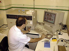
Diagram of cytotoxicity studies and their different forms of induction: by CD 8+ lymphocytes, by NK cells and by complement-activating immunoglobulins. Their evaluation is carried out by measuring chromium 51 liberation in a g radioactive particle counter after destroying the target cell.

CYTOTOXIC ACTIVITY OF T LYMPHOCYTES (CD 8+)

The cytotoxic activity of T lymphocytes (CD 8+) against a target cell can be studied by measuring the capacity that a certain number of T lymphocytes have to destroy a certain number of target cells when both populations are in contact. There are several methods to evaluate the percentage of lysis or cell death in the target cells. The most-commonly method, because of its precision, sensitivity and reproducibility, is that of chromium 51 liberation from target cells. In short, this method consists of the following: The target cells (which express certain membrane antigens) are labelled with chromium 51 and put in contact with the effector cells (CD 8, NK, etc) in a suitable proportion. After the incubation period, both cell populations are centrifuged and then a portion of the resulting supernatant is analyzed using a gamma particle counter to find out the percentage of chromium 51 released. The greater the quantity of chromium released, the greater the cytotoxic activity.

Cells of the porcine immune system | Characteristics of porcine lymphocytes | Other lymphocytes | How are the cells studied? | Swine histocompatibility antigens. (SLA) | Self-evaluation
© 2006. José Manuel Sánchez-Vizcaíno.
Design and Website by Interbionet.
Meet the author