Introductory Course in Swine Immunology. 2nd ed.
Chapter 2. Cells of the porcine immune system.
 
 
Porcine histocompatibility antigens.

All mammals possess a collection of genes which follow Mendel’s laws. They code proteins which are expressed in the cell membrane. Their polymorphism determines the acceptance or rejection of a graft. These genes help the immune system differentiate between its own and foreign molecules.

This cluster of genes is called Major Histocompatibility Complex or MHC. These genes play a key role not only in the immune response against grafts or transplants, but also in the control of antigen presentation and in the development of the immune response. The molecules coded by the MHC genes bind to the foreign proteins. They are labelled so that the immune system can recognise them and act against them.

DISTRIBUTION OF PORCINE SLA.

SLA I: All nucleus-containing cells
SLA II: Monocytes, macrophages, dendritic cells, B lymphocytes
SLA III: They are not found in cells

All porcine cells, except for the erythrocytes, have proteins on their surface whose polymorphism determines whether there is rejection or not of organs transplanted between two different animals. In pigs these major histocompatibility complex (MHC) proteins are called Swine Leucocyte Antigens or SLA. They were described for the first time at the beginning of the 1970s, when a correlation was noted between acute transplant rejection and a group of antigens expressed in the surface of peripheral lymphocytes. The genes responsible for the coding of the SLA are found in chromosome 7 and have an approximate size of 2 Mb. Around 70 genes have already been classified. They are linked to the gene coding systems J and C. The same as in other animal species, three histocompatibility antigens have been found in pigs in function of their chemical structure, their tissue distribution and their function. They are named:

SLA I: SLA II: SLA III.

Class I and III antigens are separated from those of class II by the centromere of chromosome 7, presenting a spatial relationship similar to that of human MHC.

Class I and III antigens.

Class I and II antigens are proteins integrated within the cellular membrane. Their structure is as follows: an extracellular region, a segment that crosses the membrane and a small portion which penetrates the cytoplasm.  

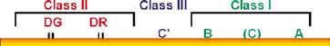
Class I antigens are expressed on the surface of all nucleus-containing cells with the single exception of the neurons and trophoblasts. As they are formed in the infected cell (viral infection), they bind to viral proteins (antigens) which are being synthesized, forming a SLA I - Antigen complex which is expressed in the membrane of the infected cell. This complex is recognised by a particular T lymphocyte, the CD 8+ or cytotoxic T lymphocyte, which kills the infected cell. Antigen presentation to CD8+ in infected cells is one of the main functions of SLA I.

Class I antigens are made of a heterodimer made up of two chains: a heavy chain called A with a molecular weight of 45 kd which is extremely polymorphic and is coded by SLA genes. The other chain, called B, is light with a lower molecular weight (12 kd). It is not coded for SLA genes and is not polymorphic. Each SLA haplotype codes 2 or 3 class I loci, named, as in the human species: A, B and C, with a total of between 7 and 10 different genes. The functional differences of the class I genes can be defined serologically, with more than 40 different alleles of SLA I having been identified.

In pigs, the class II antigens are expressed in a more restricted manner. They are found in the B lymphocytes, antigen-presenting cells and in several subpopulations of T lymphocytes, irrespective of their state of activation (this is typical of the porcine species). SLA II also has the function of presenting antigens to the CD4+ T lymphocytes, but in this case by means of phagocytic or antigen-presenting cells which process the antigens by enzyme degradation and not by cellular infection as is the case of SLA I. The molecules captured by these cells are degraded into small particles, associated to SLA and expressed in the cell membrane, forming a SLA II - Antigen complex. In this case the T lymphocyte which recognises this complex is the CD4 or T helper cell. The B lymphocyte also expresses SLA II. (Chapter 3)

SLA II genes

1 SLA-DRA

2 SLA-DRB

1 or 2 SLA-DQA

2 or 3 SLA-DQA

1 SLA-DPA

1 SLA-DPB


The class II antigens are represented by the loci: SLA-DR and SLA-DQ. The class II antigens are made up of two chains of glycoproteins known as a and b which present a molecular weight of: 33 to 35 kb in chain a and from 27 to 29 in chain b. Both chains are coded by genes of SLA a and b which presents a molecular weight of: 33 to 35 kb in chain A and 27 to 29 in chain B. The SLA family comprises around different 10 genes.  

The SLA III codes:

Complement factors: C4 and C2
21-Hydroxilase
Tumoural Necrosis Factor

Class III genes: In contrast to SLA I and SLA II, they code proteins that are not found on the cell surface but instead in the blood. Therefore some of the complement components are coded by SLA III, also being involved in other less specific actions of the immune system, such as the selection of tumoural necrosis factors.

The SLA antigens regulate the immune response against a high number of pathogens, playing a key role in antigen-presenting mechanisms.

THE RECOGNITION OF ANTIGENS BY LYMPHOCYTES IS CARRIED OUT BY ASSOCIATION WITH SLA.

Cells of the porcine immune system | Characteristics of porcine lymphocytes | Other lymphocytes | How are the cells studied? | Swine histocompatibility antigens. (SLA) | Self-evaluation
© 2006. José Manuel Sánchez-Vizcaíno.
Design and Website by Interbionet.
Meet the author