|
|||||
|
|
SWINE
LEUCOCYTE ANTIGENS (SLA) |
||||
|
|||||
|
|
SWINE
LEUCOCYTE ANTIGENS (SLA) |
||||
Every mammal posseses a cluster of genes that follow the Mendel Laws. They encode cell surface proteins. Their polymorphism determines graft acceptation or rejection. These genes help the immune system to differentiate between its own and foreign components. |
||||||||
|
SLA DISTRIBUTION.
|
This gene cluster is known as Major Histocompatibility Complex (MHC). It plays a main role, not only in the immune response rejection of tissue grafts, but also in the antigen presentation and immune response development. Molecules codified by MHC genes bind to foreign proteins, labeling them so that the immune system can recognize them and act against them. |
|||||||
|
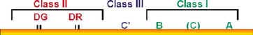
Every swine cell, with the exception of erythrocytes, have some surface proteins whose polymorphism determine the rejection or acceptation of grafted tissues between two different individuals. The proteins of the Major Histocompatibility Complex (MHC) of the pig are known as SLA (Swine Leucocyte Antigens). They were first described in the seventies, when a correlation was found between acute graft rejection and a group of surface antigens present on peripheral lymphocytes. The genes encoding SLA are located in the chromosome 7, and have an approximate size of 2 Mb. 70 of these genes have already been described. They are related to gene encoding systems J and C. As in other animal species, pigs have three different histocompatibility antigens. They are desctribed according to their: chemical structure, tissue distribution, and their function. They are known as: |
||||||||
|
||||||||
|
|
||||||||
|
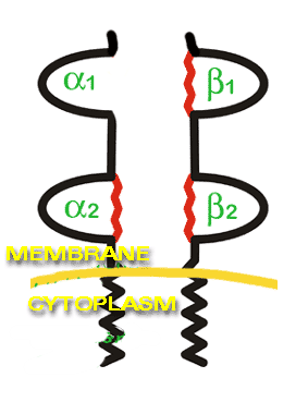
Antigens of classes I and II are proteins integrated in the cell membrane. Their structure is as follows: an extra cellular region, a segment inserted in the membrane and a small portion inside the cytoplasm.
Antigens of classes I and III are separated from those of class II by the centromere of chromosome 7, and they have a similar location to that of the human MHC. |
|
|||||||
|
Class I antigens are located in every nucleated cell surface, excepting
neurons and trophoblasts. While they become synthesized in the infected cell (viral infection),
they bind to the viral proteins giving rise to a SLA I- antigen complex. This
complex will be recognized by certain T lymphocytes, CD 8+ cytotoxic T cells, that kill the infected
cells. Antigen presentation in infected cells to CD8+ is one of the main
functions of SLA I. |
Class I antigens consist of two chains: a heavy chain, of 45 Kd, with a great polymorphism. It is codified by SLA genes and known as the A chain. The other chain is a light chain, with a molecular weight of 12 Kd, called B. The light chain is not codified by SLA genes and is not polymorphic. Each SLA haplotype codifies 2 or 3 class I loci, known, as in the human species, as A, B and C, with a total number of genes of 7 to 10. Functional differences of class I genes can be serologically defined, and more than 40 different alleles of SLA I have been identified. |
|||||||
|
Class II Antigens are expressed in the pig in a more restricted way. They are located in B lymphocytes, antigen presenting cells, and several T lymphocyte subpopulations. These T lymphocytes express class II antigens independently of their stage of activation, which is typical in the porcine species. SLA also has the function of presenting antigens to CD4+ cytotoxic T cells, but in this case, by means of phagocytes or antigen presenting cells. These cells process antigens by enzyme degradation, and not by cell infection as in the case of SLA I. Captured molecules are reduced to small fragments, associated to SLA and expressed in the cell surface, forming a SLA II- Antigen complex. In this case, the T lymphocyte that will recognize this complex are CD4 T cell or a helper T lymphocyte. B lymphocytes also express SLA II (chapter 3). |
||||||||
|
Class II antigens are codified by loci: SLA-DR and SLA-DQ. Class II antigens consist of two glycoprotein chains named a and b. a chain has a molecular weight of 33 to 35 kd and b chain of 27 to 29 kd. The SLA family is made up of some 10 different genes. |
SLA II genes
|
|||||||
SLA III codifies:
. |
Genes codifying for class III: In contrast with SLA I and II, they codify proteins that are not in cell
surface, but in the blood. Thus, some of the complement
elements are codified by SLA III. They play a role in other less specific immune actions, such as the
selection of tumor necrosis factors. |
|||||||
| SLA
antigens regulate the immune response against a large number of pathogens,
and play a major role
in antigen presentation mechanisms. |
||||||||
| LYMPHOCYTE RECOGNITION OF ANTIGENS IS DONE BY SLA ASSOCIATION. |
||||||||
|
|||||