|
|||||
|
|
HOW ARE
CELLS STUDIED? |
||||
|
|||||
|
|
HOW ARE
CELLS STUDIED? |
||||
|
As was indicated at the beginning of this chapter, it is not possible to differentiate between B and T lymphocytes using ordinary microscopy, either in blood or in lymphoid organs. However, some differences are observed when electron microscopy is used. Nevertheless, there are some methods that allow differentiation and quantification of lymphocytes, their subpopulations, and even their capability to respond to different antigens. The more frequently used methods are grouped as follows: |
||||||||
|
||||||||
|
For B lymphocytes: B lymphocytes have in their membrane surface
immunoglobulins that can be detected using anti-porcine-immunoglobulin sera or immunoglobulin.
These anti-immunoglobulin antibodies can be labeled with fluorescent dyes; monoclonal antibodies
labeled with fluorescein isocyanate can also be used.
All of them are specific against each immunoglobulin isotype. Using a fluorescence microscopy, a positive fluorescent reaction in
the membrane can be observed . |
||||||||
|
|
|
|||||||
|
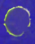
Surface immunoglobulins of a B lymphocyte observed by fluorescence microscopy. |
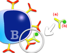
Technique used for the observation of surface immunoglobulins of B lymphocytes. A polyclonal or monoclonal serum (a), labeled with fluorescein isocyanate (b), reacts with porcine immunoglobulins of the membrane. |
|||||||
|
For T lymphocytes. Rosettes formed with
erythrocytes. Porcine T lymphocytes, like those of other species, have the characteristic of forming
E-rosettes when they bind selectively to sheep red blood cells (a). Lymphocytes can be quantified
by acridine orange labeling and
afterwards observed with a fluorescence and ordinary light microscope. If T and B lymphocytes are
labeled with acridine orange, only T lymphocytes form E-rosettes; B lymphocytes are
actually stained but do not form rosettes.
|
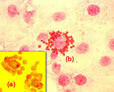
It is important to note that other
rosette types may be formed by porcine cells. In the above picture
rosette
formation of porcine erythrocytescan be observed in macrophages infected by the African Swine Fever virus (b).
This phenomenon is known as hemadsorption and must not be confused with E-rosette formation with sheep
erythrocytes by porcine T lymphocytes (a). |
|||||||
The different porcine
lymphocyte populations can be labeled using monoclonal antibodies. The more frequently used methods
are: |
||||||||
|
||||||||
|
2.1. Flow cytometry. A flow cytometer allows "in vitro" characterization and even separation of the different lymphocyte populations by means of monoclonal antibodies labeled with fluoresceine, these antibodies are specific for the surface markers of each targeted population. Nowadays, flow cytometry allows the evaluation of several cytocromes at the same time. Thus, different cell subpopulations can be studied in the same lymphocyte sample. It is possible to start with whole blood (state of the art cytometers allow working with whole blood) or with the lymphocyte population previously separated from the blood. The cells, with the specific monoclonal antibody (or antibodies; may be several at the same time) against the marker under study, are forced through a nozzle in a single cell stream that passes through a laser beam. This detects the scattering of light (particle size) and emitted fluorescence (cell type). The obtained measurements indicate each population percentage. |
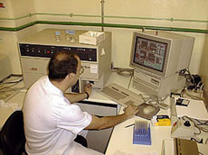
Picture of one of the flow cytometers used for the study of porcine lymphocyte populations. The basic lecture unit and data processing system can be observed. |
|||||||
|
|
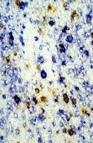
2.2. Immunohistochemistry. Using monoclonal antibodies, labeled with fluoresceine or peroxidase, against the different porcine lymphocytes, it is possible to study these cells in any tissue. It is even possible to study two different populations at the same time. This is achieved using two monoclonal antibodies against two different lymphocyte populations. Each of them is labeled with a different enzyme (double labeling) (peroxidase- alkaline phosphatase). These techniques have been of great importance in the study of the pathogenic mechanisms in several porcine infectious diseases. Thus, in the case of African Swine Fever Virus (ASFV), the different lymphocyte populations affected, or not, by the virus were studied. It was observed that those macrophages which were infected by the virus, had an altered SLA expression. Nowadays, these techniques are used to know the affected populations in the different viral infections and to study the different pathogenic mechanisms. |
|||||||
|
Double labeling study (brown-blue) for the localization of ASFV infection (brown) in cells labeled with a monoclonal antibody against porcine macrophage (blue). No all cells reacting with the monoclonal antibody (blue) are infected, although most of them are. |
||||||||
|
These methods are based on the evaluation of the B and T lymphocytes' capability to recognize a specific antigen. The most currently used techniques are:
|
||||||||
|
INDUCED LYMPHOCYTE PROLIFERATION.
|
||||||||
|
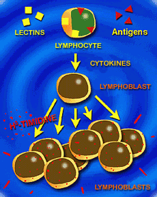
Induced lymphocyte proliferation. The use of lymphocyte transformation or blastogenesis is nowadays one of the more precise and most frequently used "in vitro" techniques for the study of the specific and non-specific stimulation capability of lymphocytes. This technique is based on the capability of the lymphocytes for responding to an antigen (specific response) which has induced memory lymphocytes, either by vaccination or by natural infection. These lymphocytes, when in repeated contact with the antigen, have a blastogenic transformation. This blastogenic transformation may be induced in a non-specific way, due to the lymphocyte's capability of reacting to different lectins or mitogens. Lectins induce a non-specific stimulation both in B and T lymphocytes. |
|
|||||||
|
Induced lymphocyte proliferation. |
||||||||
|
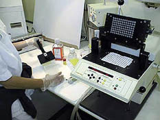
Picture of the equipment used for harvesting cells cultured in plaques. It must be noted that cells remain in the filter paper. These filters will later be analyzed using the liquid scintillation counter or b particle counter. |
|
|||||||
|
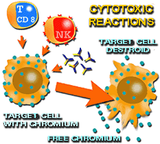
Cytotoxicity study, taking into account
the different ways of induction: by CD 8+ lymphocytes, by NK cells and by immunoglobulins
that activate complement. Their evaluation is carried out by measuring Chromium 51 liberation after
the destruction of target cells, using a g racioactive particles counter . |
CYTOTOXIC ACTIVITY OF T LYMPHOCYTES (CD 8+) The cytotoxic activity of T lymphocytes (CD 8+) towards a target cell can be studied measuring the killing ability that a determined number of T lymphocytes have for a certain number of target cells, when both populations are placed together. There are several methods for the evaluation of lysis percentage or target cells killed. However, the one most used is the liberation of Chromium 51 coming from target cells. In this method, target cells (which express certain surface antigens) are labeled with Chromium 51, and then placed together with effector cells (T lymphocytes). After the incubation period, cells are centrifuged and then, chromium 51 in the collected supernatant is measured, using a gamma-particle counter. The more chromium present in the supernatant, the larger the cytotoxic activity. |
|
|||||