|
|
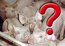
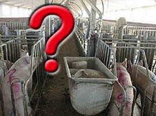
WHAT IS A SERUM PROFILE?
|
|
|
|
WHAT IS A SERUM PROFILE?
|
| Serum
profiles are those serial studies performed in order to know the immunological and sanitary status of
a farm. |
|||||||||||||||||||||||||||||
|
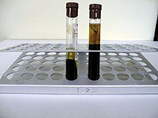
Serum profiles are based on the detection of circulating antibodies. This can be done by using any of the techniques now available, in order to obtain the necessary information about the immunological status of an animal at that specific moment or in previous stages. It is slowly becoming more common to use these studies in the sanitary control of pig farms. They are, in many cases, an important tool for reducing expenses and increasing the sanitary level. Serological studies allow one to, among other things: know the sanitary status, choose the best time for vaccination, control vaccination programs and avoid the introduction of new diseases to the farm. |
|
||||||||||||||||||||||||||||
|
We must
take the following factors into account in order to perform a good
serologic study of a farm:
|
|||||||||||||||||||||||||||||
|
|
||||||||||||||||||||||||||||
|
|||||||||||||||||||||||||||||
|
|
|
||||||||||||||||||||||||||||
|
From the moment the piglet sucks colostrum it will passively acquire humoral immunity. This immunity will be similar to the mother's. The maximum intake takes place between 6 to 36 hours after birth, and we can detect immunoglobulins in the bloodstream between 12 and 24 hours after colostrum ingestion. The length of time these antibodies remain in the blood stream is variable and will depend on the type of immunoglobulin, the mother's immunological status, the infectious agent, etc. Usually colostrum immunoglobulins can be found in weeks 3, 8 and 20. This is one reason among others as to why it is important to know at what age we should take the sample from the animal and from which age group it should be taken, i.e.; sows, piglets, etc.
|
|||||||||||||||||||||||||||||
|
|||||||||||||||||||||||||||||
|
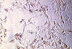
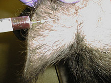
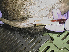
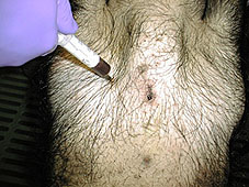
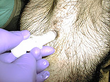
Blood used for serum collection is usually obtained from: Ear marginal vein. Using a lancet and collecting the blood in a tube or using syringes with 16 or 25 mm gauge needles. This system has the advantage of being easy to perform, but the amount of blood collected is small, and contaminated samples are frequent. Tail vein. Usually with syringes (25 mm gauge needles), vacutainers or partial amputation. This method is also easy to perform, but the sample size obtained is only about 0.5 ml. It is not the ideal method. Jugular vein. Is one of the most frequently used methods, especially in sows. Usually with syringes, vacutainers, or monovet. The amount obtained is about 10 to 30 ml. Cava vein. Used to bleed all type of animals,
from piglets to adults, and specially when a large amount of blood is needed. Needles vary
depending on the size of the animal (10 Kg: 25 mm; 45 Kg: 38 mm; 100 Kg: 50 mm; sows of 100 Kg: 100
mm)
|
|
||||||||||||||||||||||||||||
|
|
|||||||||||||||||||||||||||||
|
|
|||||||||||||||||||||||||||||
|
|
|||||||||||||||||||||||||||||
|
|
|||||||||||||||||||||||||||||
As we mentioned at the beginning of this chapter,
there are several laboratory techniques which are available nowadays for serological studies.
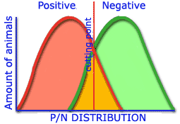
The diagnostic capability of a technique is determined by the evaluation of its sensitivity and
specificity compared to the reference technique. Some concepts must not be forgotten when making
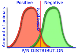
the assessment of the available techniques. These are: Specificity: Is the method's capability to discriminate between positive and negative sera. A good specificity must lack false positive cases. No negative case must be considered as positive by any technique. Predictive value: Is the capability of the technique to discriminate between animals that are suffering a given disease and animals which are not. In other words, it predicts the sensitivity and specificity for either a negative or a positive case. The predictive value can be:
Efficacy: Percentage of correctly classified animals. True positive: Sick animal that has been correctly classified by the technique. False positive: Animal that has not been correctly classified by the technique. |
|||||||||||||||||||||||||||||
|
The assessment of the sensitivity, specificity, and predictive value of any technique is performed by comparing its results with those obtained with a technique of reference or with the real disease, using the following formula:
A: Positives for both techniques. B: Real positives that are considered as negative by the assessed technique. C: Positives of the assessed technique that are real negatives. D: Negatives for both techniques.
Predictive value for negatives: |
|
||||||||||||||||||||||||||||
|
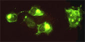
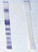
Among the techniques that are currently being used, those with better sensitivity and specificity levels are seroneutralization and ELISA. Depending on the laboratory, similar results can be obtained using both techniques. The most important thing is to remember that if we want to compare results at different times, results at different stages... the same technique should be used, and even the same laboratory. If not, different results could be obtained and the diagnostic would not be correct.
|
|||||||||||||||||||||||||||||
|