|
|
HOW ARE IMMUNOGLOBULINS STUDIED?
|
|
|
|
HOW ARE IMMUNOGLOBULINS STUDIED?
|
|
Porcine immunoglobulins can be
found in two different forms: as part of the BcR receptors of B lymphocytes or as antibodies in the
serum, body fluids and tissues. Their study is a basic key for Animal Health. |
||||||||||||||||||||||||
|
As we have already shown in chapter 2, How are the B lymphocytes studied?, there are several ways of studying surface immunoglobulins as part of the BcR receptors of B lymphocytes. The two most frequent are:
Those immunoglobulins found as
free molecules in serum, colostrum, milk, fluids, etc. can be studied both as total immunoglobulins,
without taking into account against which antigen they can react to, and secondly, as specific
antibodies against different antigens of interest. A
huge number of available techniques are used in this last type of study,
which is without a doubt the
most interesting. They range from the classic precipitation or complement activation to the most modern and gradually more
used immuno-enzymatic techniques. |
|
|||||||||||||||||||||||
|
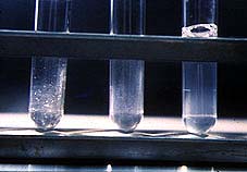
Differences in sensitivity, specifity, and the possibility of use in a large number of samples, etc. are some of the most important concepts that must be taken into account when choosing one of these techniques. The sensitivity level for example is low in techniques such as precipitation. Bacterial agglutination and complement activation have an average sensitivity.Finally, techniques such as ELISA, seroneutralization or bactericide activity have a greater sensitivity.
Image showing a tube agglutination The development of Molecular
Biology and the production of monoclonal
antibodies have allowed us, in the last years,
to have diagnostic tools with
a great sensitivity and specificity. They are sometimes even presented as KITS,
which are both easy and simple to use and
read. |
|
|||||||||||||||||||||||
|
Among the methods available now, we can point out, those that have more possibilities of performing serologic studies on a large scale level, and without the need of highly technical resources. We will also study other methods that permit us to study only a small number of samples, but have good sensitivity and specifity and do not require expensive equipment. Finally, we will see a technique of reference for every serologic study, which needs a larger infrastructure and longer performance time, but due to its great importance, we have included it in this chapter. |
||||||||||||||||||||||||
The
main methods with these characteristics are:
|
||||||||||||||||||||||||
|
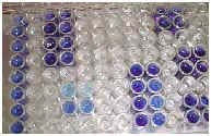
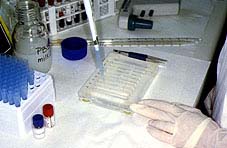
The enzyme-linked immunosorbent assay (ELISA) is one of those serological techniques that can detect the ANTIGEN-ANTIBODY reaction by the use of enzyme-linked antibodies . ELISA is based on the use of labeled antibodies (usually done with the enzyme peroxidase) so that the resulting conjugates have both immunological and enzymatic activity. One of the components of the conjugate (antibody or antigen) is attached to the plate, so the antigen-antibody reaction can be easily measured when adding the reaction substrate. This substrate produces a colored reaction product when it comes into contact with the enzyme. The color change can be seen or quantified with a colorimeter. There are several commercial KITS available on the market, so serological studies can be performed without the need of large infrastructures.
|
We can observe the positive control serum in the first row and the negative control serum in the second row. The unknown samples are in the rest of the wells. Positive samples are blue and the negative ones are colorless. |
|||||||||||||||||||||||
|
Several types of ELISA have been adapted both for the detection of ANTIGENS and ANTIBODIES: |
||||||||||||||||||||||||
|
The most frequent ELISA for antigen detection is the Sandwich ELISA. In this type of ELISA, plates are usually coated with an antibody (monoclonal or polyclonal antibody) against the unknown antigen. A macerate of the organ that needs to be tested is added to the wells, and if the antigen is present, the antigen-antibody reaction will take place. After that, we add another antibody linked to an enzyme. When the reaction substrate is added it turns a color. |
Steps of a Sandwich Elisa. (1) A monoclonal or polyclonal antibody is usually attached to the plate. (2) Incubation with the unknown sample. (3) Addition of the conjugate. (4) Substrate addition. Every step is preceded by incubations and washings. |
|||||||||||||||||||||||
|
Usually the following types of ELISA are used for the detection of specific antibodies:
|
||||||||||||||||||||||||
|
Indirect ELISA steps: (1) Antigen is bound to the plate. (2) Addition of test serum. (3) Addition of the conjugate. (4) Addition of the substrate. |
It is the most commonly used method for antibody
detection. Briefly, it involves the coating of the ELISA plate with the antigen (kits have
plates already coated) against the specific antibodies that may be present in the serum. Antigens can
be viral or bacterial proteins, and or even whole virus molecules.
|
|||||||||||||||||||||||
|
This technique is also very common for the detection of specific antibodies. We have an antibody (monoclonal of polyclonal) of a known antigen. This antigen has previously been bound to the plate. It is known as competitive ELISA because the serum is incubated with the antigen previous to its incubation with the antiserum bound to the plate. Therefore, both compete for the antigen. The following steps include addition of the conjugate, incubation, washing, and finally, substrate addition and reading the results.
|
|
|||||||||||||||||||||||
|
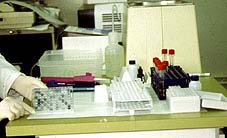
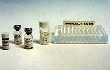
Required material to perform
Immunoelectrotransference Technique:
antigen-nitrocellulose sheets, PBS tampon, positive and negative control sera, conjugate, substrate
solution and plastic plate. |
IMMUNOELECTROTRANSFERENCE OR WESTERN BLOT Immunoelectrotransference, "western blot" or "Immunoblotting" is an immunoenzymatic technique used for the detection of specific antibodies. It is recommended whenever it is necessary to study a large number of sera which have given doubtful results using other techniques. This technique uses nitrocellulose sheets as an antigenic substrate, to which antigenic proteins have previously been transferred maintaining completely or in part their antigenic characteristics. |
|||||||||||||||||||||||
|
In order to obtain the antigen-nitrocellulose sheets, proteins are first separated
by polyacrylamide gel electrophoresis (SDS-PAGE). Later, these proteins are electrically transferred
from the gel to the nitrocellulose sheets. These sheets are then cut and will become the antigen substrate.
Each one of these pieces are then incubated with the test sera and washed. Then, a labeled
anti-immunoglobulin (IgG or IgM) is added. If there is any immunoglobulin bound to the antigenic
protein, they will be revealed by the addition of the conjugate. One or more specific precipitation
lines will be observed depending on the existence of specific antibodies against one or more proteins.
Specifity is detected when immunoglobulins react with proteins of the same molecular weight as
that of the antigenic proteins. |
||||||||||||||||||||||||
|
It is a very sensitive technique, easy to perform and to interpret. No special equipment is needed. This technique is especially indicated for the study of small numbers of sera. As it does not require special equipment, it is possible to perform it in laboratories with little equipment. Nowadays, there are several commercial kits available, but more kinds of kits will eventually become available.
|
The last step of the technique. It is possible to observe the different lines where test and control serum have reacted. |
|||||||||||||||||||||||
|
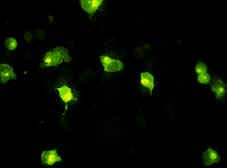
Indirect immunofluorescence technique. MS cells infected by the African Swine Fever virus. Antibodies bound to the infected cells can be observed, those areas of the cytoplasm with higher viral replication have more bound antibodies and therefore, a higher light intensity. |
INDIRECT IMMUNOFLUORESCENCE OR IMMUNOPEROXIDASE Indirect immunofluorescence or immunoperoxidase
(depending on the use of either isocianate or peroxidase), are techniques that use the specifity of
histology (cells can actually be seen and the location of the immunological reaction can be
determined) and the sensitivity of the immunological techniques.
|
|||||||||||||||||||||||
|
This method is considered as the reference technique for every serological study. This is due to the fact that results obtained from it will correlate the best with "in vivo" responses. The use of this test has made it possible to measure the capability of antibodies present in the test sera of neutralizing the biological activity of an antigen. The methods described above allow the evaluation of a serum for recognizing an antigen. In seroneutralization, we go a step further, and the potential of the serum of neutralizing the biological activity of an antigen can also be known . These methods are very common in laboratories when the assessment of the capability of a serum against bacterial toxins, bacteria or viruses is needed. They are more laborious, need cell cultures, sterile conditions, and usually require more time than those studied above. They are however, highly specific and sensitive and are considered as reference methods for every serological evaluation. |
||||||||||||||||||||||||
|
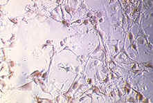
Infected cell layer. |
In the case of viruses, we can measure the capability of a given serum for neutralizing the virus infectivity on a susceptible cell line. A viral solution, of a constant concentration and which has previously been in contact with different dilutions of the test serum, is added to the cell culture. The observation of the cells at different times allows one to see if these cells are being infected or not by the virus, using either conjugated dyes or looking for the cytopatic effect. We can measure, in this way, the serum capability for neutralizing the virus.
|
|||||||||||||||||||||||
|
© Copyright. 2001. José Manuel
Sánchez-Vizcaíno Rodríguez. All rights reserved.
Dep. Legal: B-32.422-01. ISBN: 84-699-5917-4