|
|||||
|
|
How do cytokines intervene in the natural or innate immune response?
|
||||
|
|||||
|
|
How do cytokines intervene in the natural or innate immune response?
|
||||
|
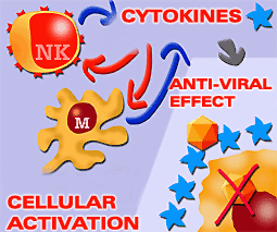
Cytokines play a main role in the natural or innate response by means of the direct action of mechanisms against the invading agent during the early stages of the infection, or by means of immune-modulatory mechanisms which activate NK cells and monocytes-macrophages; which then induce the release of cytokines.
|
||||||||||||||||||||||||||||||||||||||
|
If any antigen gets through the chemical and physical barriers, innate immunity provides a first line of defense. The innate immunity system provides the first non-specific defense barrier of the pig. In this type of response cytokines play a very important role, both directly (for example, blocking viral replication by the interferons) and by means of different immune-modulatory mechanisms that trigger the inflammatory response, produce and elevation on the body temperature, activate NK cells and macrophages, etc. Those cytokines that have a role in this step are mainly produced by monocytes-macrophages and other non-immunological cells, such as fibroblasts and endothelial cells.
|
Cytokines play a main role in the innate immune response by means of direct mechanisms against the invading agent (inhibiting viral replication) or by activating mechanisms for cells such as NK cells and macrophages, which upon activation, produce more cytokines. |
|||||||||||||||||||||||||||||||||||||
|
The main cytokines that have a role in the innate response are:
|
The IL 1, IL 6, IL 12 intervene in the natural response by activating the monocytes-macrophages and NK cells, and by allowing the activation of the mechanisms responsible for the elevation of the body temperature. They activate the immune response and reduce the capability of replication of the pathogen.
Cytokine IL 16 seems to be related to the activation of T CD 4+ lymphocytes. T CD 4+ |
|||||||||||||||||||||||||||||||||||||
|
CHARACTERISTICS OF CYTOKINES IMPLICATED IN THE INNATE RESPONSE.
|
||||||||||||||||||||||||||||||||||||||
|
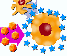
Interferon (IFN) can be a, b and g, and acts in different ways in order to inhibit viral replication. The first of them is induced by IFN a and b, which are mainly produced by monocytes-macrophages, and in a lesser proportion by fibroblasts. It consists of the production of a transitory stage of resistance against viruses in those cells susceptible to infection by the virus. This effect, with a great anti-viral capability, does not need large quantities of interferon, and is one of the main mechanisms of the innate immune response. |
Transitory resistance |
|||||||||||||||||||||||||||||||||||||
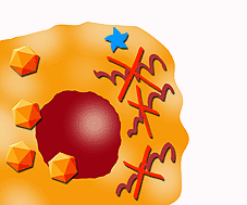
| IFN also induces the synthesis of certain molecules with anti-viral properties, such as oligoadenilate-synthetasa, which interferes with DNA and RNA or protein synthesis, due to different enzymes which, in the presence of double-stranded RNA, activate an endoribonuclease that then degrades viral mRNA. |
mRNA degradation. Protein synthesis inhibition. |
|||||||||||||||||||||||||||||||||||||
|
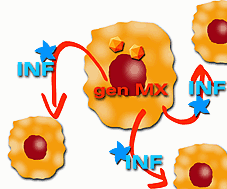
Lastly, IFN a and b are thought to activate several genes that express anti-viral proteins, such as the Mx gene, that changes murine influenza susceptible cells into resistant ones. In these mechanisms, infected cells are able to secrete interferon that protects uninfected adjacent cells.
In addition, interferons increase the expression of SLA I in the infected cells, which allows the recognition of this cells by CD 8+ lymphocytes and NK cells, increasing the cytotoxic activity.
|
Activation of genes that express anti-viral proteins. |
|||||||||||||||||||||||||||||||||||||
|
Increase in SLA expression |
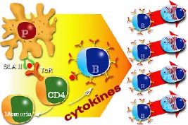
IFN g has a different structure from IFN a and b. IFN g is produced mainly by T CD4+ and CD8+ lymphocytes and by NK cells after an antigenic stimulation. Besides its anti-viral activity, IFN g plays a role in many immune-modulatory functions, such as the increase of the expression of SLA I (which will enhance the cytotoxic activity) and SLA II (which will favor cell cooperation in antigen presentation and antibody production). |
|||||||||||||||||||||||||||||||||||||
|
Cell cooperation for antigen presentation and the production of antibodies. Cytokines play a main role in these processes. |
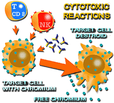
By cytotoxicity techniques using Chromo 51 liberation it is possible to make the assessment of cytotoxic effects in vitro. |
|||||||||||||||||||||||||||||||||||||
|
Interferon has been detected in pigs during infections by several viruses (Transmissible Gastroenteritis, Influenza, Classic Porcine Fever, Aujeszky´s), and in some of these diseases, its capability to increase or modulate SLA expression has been demonstrated. |
||||||||||||||||||||||||||||||||||||||
|
IFN-g is one of the cytokines that has been cloned in many species, and a large number of studies have been performed on its homology and cross reactivity. One can conclude from these studies that that IFN-g can be classified in three groups depending on functional and structural homology. The first group is composed of primate and human IFN-g; the second group comprises IFN-g from bovine, ovine, deer, porcine and horses. In the third group, we find IFN-g from mice and rats. In general, most of the cytokines cloned have certain homology in respect to their sequence and so they present cross reactivity. |
||||||||||||||||||||||||||||||||||||||
| CHAPTER 6 | Previous theme | Next theme | Course program |