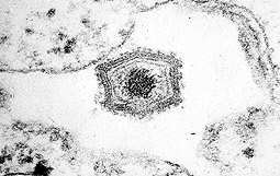
Photograph of African Swine Fever virus under electron microscope
|
Viruses need the machinery of the infected cell to synthesize their own proteins. They are intracellular parasites that depend on the infected cell for replication. Viruses generally have very simple structures consisting of proteins and nucleic acid, although their structure and composition is very varied. They are divided into two groups on the basis of the nucleic acid they carry: DNA and RNA viruses, and into different families by their form and structure.
|
|
MAIN VIRAL DISEASES OF THE PIG
|
VIRUS |
FAMILY |
NUCLEIC ACID |
|
ADENOVIRUS |
ADENOVIRIDAE |
DNA (dc) |
|
PORCINE CYTOMEGALOVIRUS |
HERPESVIRIDAE |
DNA (dc) |
|
PORCINE CIRCOVIRUS |
CIRCOVIRIDAE |
DNA (sc) |
|
PORCINE ENCEPHALOMYOCARDITIS
|
PICORNAVIRIDAE |
RNA (sc) |
|
JAPANESE ENCEPHALITIS |
FLAVIVIRIDAE |
RNA (sc) |
|
AUJESZKY'S DISEASE |
HERPESVIRIDAE |
DNA (dc) |
|
BLUE EYE DISEASE |
PARAMYXOVIDAE |
RNA (sc) |
|
SWINE VESICULAR DISEASE |
PICORNAVIRIDAE |
RNA (sc) |
|
PORCINE ENTEROVIRUS |
PICORNAVIRIDAE |
RNA (sc) |
|
VESICULAR STOMATITIS |
RHABDOVIRIDAE |
RNA (sc) |
|
VESICULAR EXANTHEMA |
CALICIVIRIDAE |
RNA (sc) |
|
FOOT-AND-MOUTH DISEASE |
PICORNAVIRIDAE |
RNA (sc) |
|
TRANSMISSIBLE PORCINE GASTROENTERITIS |
CORONAVIRUS CORONAVIRIDAE |
RNA (sc)
RNA (sc) |
|
PORCINE INFLUENZA |
ORTHOMYXOVIRIDAE |
RNA (sc) |
|
PORCINE PARVOVIRUS |
PARVOVIRIDAE |
DNA (sc) |
|
AFRICAN SWINE FEVER |
Family without name
(genus ASF -Like Virus) |
DNA (dc) |
|
CLASSIC SWINE FEVER |
FLAVIVIRIDAE |
RNA (sc) |
|
PORCINE ROTAVIRUS |
REOVIRIDAE |
RNA (dc) |
|
PORCINE REPRODUCTIVE AND RESPIRATORY SYNDROME |
ARTERIVIRIDAE |
RNA (sc) |
(sc): single chain. (dc): double chain |
|
From the immunological point of view, it is of interest to discover the viral replication cycle, in order to foresee the opportunities available to the different immune mechanisms to interact with the viral particle, with infected cells, or with both. Normally, the viral replication cycle starts by the binding of the virus (free virus) to the host cell via specific receptors (adsorption) (1These receptors mark the tropism and specificity of an infection (they cannot infect every cell or every species: they have their own specific tropisms). Once in the cell, the virus removes its envelope, freeing its nucleic acid (uncoating) (2), and the viral replication process begins. |
|
Diagram of the viral replication cycle. (1) Adsorption (2) Uncoating (3) Replication and (4) Assembly and viral release.
|
In this phase, cell protein synthesis is inhibited and only the genetic information of the virus will be processed. The mechanisms that act in this phase depend on the type of nucleic acid of the virus (DNA or RNA). In the In the case of the DNA virus, replication takes place (3) forming a new viral DNA. The new viral DNA is transcribed (transcription) into viral RNA (blue), which through translation creates the different viral proteins and then viral assembly (4). In the case of the RNA virus, transcription is not necessary, and the new viral RNA passes directly to protein production. This RNA replication mechanism is different for retroviruses. By means of inverse transcriptase, viral DNA is formed from the viral RNA (this binds to the cell genome), enabling the replication phases, etc to begin.
In this phase, cell protein synthesis is inhibited and only the genetic information of the virus will be processed. The mechanisms that act in this phase depend on the type of nucleic acid of the virus (DNA or RNA). In the case of the DNA virus, replication takes place (3), forming a new viral DNA. The new viral DNA is transcribed (transcription) into viral RNA (blue), which through translation creates the different viral proteins and then viral assembly (4). In the case of the RNA virus, transcription is not necessary, and the new viral RNA passes directly to protein production. This RNA replication mechanism is different for retroviruses. By means of inverse transcriptase, viral DNA is formed from the viral RNA (this binds to the cell genome), enabling the replication phases, etc to begin.
|
|
Diagram of the different immune mechanisms against viral particles (antibodies, cytokines, complement) or against the infected cell (NK, CD 8, ADCC, complement). |
In most viral infections, the immune system has the opportunity to attack the viral particles at certain times during infection (before penetrating the cell or leaving it after replication). It also attacks infected cells (in the protein production phase or in that of viral assembly), as infection antigens which appear in their membrane activate the immune response. In some cases, such as endogenous porcine retrovirus, (three types have been identified: A, B, B1 and C), or in the herpes virus (Aujeszky’s disease), the infection remains for long periods of time without the viral particle appearing, nor the infected cells expressing membrane antigens. At these times, the immune system mechanisms are ineffective, as the enemy does not offer any type of signal, but at a certain moment (not all of the circumstances are known) the infection is reactivated and goes on to free new infectious virions. |
|
Infection by retrovirus
|
From the point of view immunity, viral infections can be fought once the physical and chemical barriers have been crossed. This can be by fighting against the viral particle (virion), against the infected cells or against both, through different natural and adaptive response mechanisms.
|
|
Natural response against viruses
The most active natural response mechanisms against viral infections are mediated by interferon and by NK cell activation. These mechanisms are more directed towards infected cells. |
Activation of inductor genes of antiviral proteins
|
Interferon is a cytokine of which three types are identified, known as: a, b and g. The first two types are produced mainly by the monocytes-macrophages and in a smaller proportion by the fibroblasts, whilst interferon g is produced by CD4 and CD 8 lymphocytes and NK cells. Interferon has a high antiviral capacity, inducing different mechanisms such as: transitory resistance of cells, induction of different molecules with antiviral activity, activation of genes that express antiviral proteins increase in expression of SLA I and SLA II. |

Diagram of NK cell
|
NK cells are naturally activated against cells infected by viruses. The activation mechanism seems to be linked to alterations in SLA expression in infected cells. The NK reaction with infected cells is not based on an antigenic reaction (the NK cells do not have TcR). This cytotoxic mechanism is very effective in viral infections.
Finally, the alternative pathway of the complement also activates effective destruction of the viral particles.
|
|
Adaptive response against viruses
Adaptive immunity reacts against viral infections, both viral particles and infected cells. The most important immune mechanisms against viral particle are the antibodies, whilst cytotoxic mechanisms are the most important against infected cells. These mechanisms are mediated by cells (CD 8+) or by antibodies and cells (ADCC) or antibodies and complement (classical pathway).
|
|
Cytotoxic mechanism induced by CD 8 lymphocytes
|
Against the viral particle.
The capsid of the viral particle is formed of proteins, and is therefore very antigenic, inducing large amounts of antibodies with different activities against viruses:
Neutralizing the infection (IgG, IgM and IgA), preventing the virus from entering the cells.
Viral agglutination (IgM), reducing the number of infectious units available.
Activation of phagocytosis by forming the antigen-antibody complex and stimulating the Fc receptor of the macrophages.
Against the infected cell.
Cells infected by viruses can express viral antigens in their membrane long before viral assembly occurs, and therefore their destruction is an excellent mechanism to prevent the formation of more viruses. The adaptive response attacks infected cells by means of antibodies (ADCC system, activation of the complement by the classical pathway, activation of phagocytosis) and also by cell cytotoxicity mediated by CD 8+ lymphocytes. This is one of the most effective mechanisms against viral infections.
Cytotoxic T lymphocytes
© James A. Sullivan, Cells Alive!
(187 Kb.)
|
|
|