| |
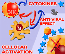
Cytokines play a key role in the natural response through direct action mechanisms against the invading agent (preventing the infection of the cells by viruses) or through cell activation mechanisms (NK and macrophages) which in turn release cytokines
|
If an antigen crosses physical and chemical barriers it sets in motion natural immunity. Natural immunity is the first non-specific immunological barrier in the pig. Cytokines play a vitally important role in this type of response. This could be direct, for example by preventing viral replication with Interferon (IFN), or by a variety of immune-regulatory mechanisms which trigger inflammation, raise the body temperature, activate NK cells and macrophages, etc. The cytokines which act during this phase are primarily produced by monocytes-macrophages and by other non-immune cells such as fibroblasts and endothelial cells.
|
|
The main cytokines involved in the natural response are:
IL 1 |
IL 6 |
IL 12 |
IL 16 |
TUMOUR NECROSIS FACTOR (TNF a) |
INTERFERON a and g |
|
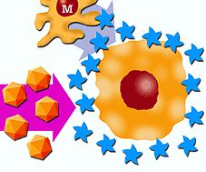
IL 1, IL 6, IL 12 are involved in the natural response by activating monocytes-macrophages and NK cells, as well as facilitating the activation of mechanisms responsible for raising the body temperature. They activate the immune system and reduce the power of multiplication / replication of the pathogen.
Tumour necrosis factor (TNF a) is the main trigger of the inflammatory response; it acts on the vessels of the affected area increasing vascular permeability, facilitating extravasation of immunoglobulins, complement, chemotactic factors, etc, helping in defence against infection.
Cytokine IL 16 seems to be linked to the activation of CD 4+ T lymphocytes
|
|
CHARACTERISTICS OF CYTOKINES RELATED TO THE NATURAL RESPONSE
| CYTOKINE |
TYPES |
C. PRODUCER |
FUNCTION |
| IL 1 |
IL 1a
IL 1b |
Macrophages
T and B lymphocytes
NK cells |
Immunological regulation.
Activation of T lymphocytes and macrophages |
| IL 2 |
IL 2a
IL 2b |
T lymphocytes (CD4)
B lymphocytes and NK cells |
Activation of T lymphocytes
Activation of macrophages |
| IL 4 |
|
Activated T lymphocytes |
Th0 - th2
SLA expression |
| IL 6 |
IL6a. IL6b |
Lymphocytes
Macrophages Fibroblasts |
Activation of T and B lymphocytes
Inflammatory |
| IL 10 |
|
T and B lymphocytes Monocytes. |
Differentiation of lymphocytes Activation of T lymphocytes Secretion of immunoglobulins |
| IL 12 |
|
Neutrophils Macrophages T and B lymphocytes |
Differentiation of lymphocytes (Th0 Th1 Th2)
Activation of NK cells |
| IL 16 |
|
T lymphocytes |
Chemotaxis |
| TNF a |
TNFaR1 TNFaR2 |
Macrophages
CD4 lymphocytes
Fibroblasts
Mast cells |
Local inflammation
Changes in permeability
Increase in phagocytosis |
| IFN a |
|
Lymphocytes |
SLA I activation |
| IFN b |
|
Fibroblasts
Other cells |
NK activation
Inhibition of virus replication |
| IFN g |
|
T lymphocytes
NK cells |
SLA I and SLA II induction
Macrophage activation |
|
|
Transitory resistance |
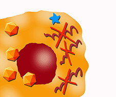
Interferon (IFN) has three types, a, b and g, which act in different ways to prevent viral infection. The first of these is induced by IFN a and b, which are mainly produced by monocytes-macrophages and to a lesser extent by fibroblasts. It consists of inducing a state of transitory resistance against infection by a whole host of viral agents in cells susceptible to infection by virus. This effect has strong anti-viral powers and does not need large amounts of Interferon. It represents one of the principal mechanisms in the natural response.
|
|
RNA messenger degradation. Protein synthesis inhibition
|
IFN also acts by the synthesis of molecules with anti-viral activity, such as oligoadenylate-synthetase, which interferes with DNA and RNA replication or inhibits cell protein synthesis, thanks to the different enzymes produced in the presence of double chains of RNA which inhibit protein synthesis through RNAse activation, which in turn degrades messenger RNA.
|
|
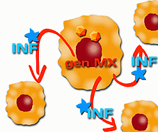
Activation of genes which express antiviral proteins |
Finally, IFN a and b are believed to activate different genes that express antiviral proteins, such as the Mx gene that changes murine influenza susceptible cells into resistant ones. In this mechanism, infected cells are able to secrete Interferon which protects non-infected neighbouring cells from infection.
These interferons also increase the expression of SLA I in infected cells, favouring their recognition by CD 8+ lymphocytes and NK cells with the resulting increase in cytotoxic activity
|
|
Increase in SLA expression
|
IFN g has a different structure to IFN a and b.
It is produced by CD4+ and CD8+ T lymphocytes and NK cells after receiving antigenic stimulation. As well as its antiviral activity, IFN g, is involved in many immune-regulatory functions, such as increasing expression of SLA I (enhances cytotoxicity) and of SLA II (favours cell cooperation in antigen presentation and antibody production).
|
|
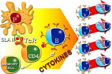
Cell cooperation for antigen presentation and antibody production. Cytokines play a key role in these processes |
|
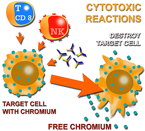
Cytotoxic mechanisms "in vitro" can be measured by cytotoxicity techniques using chromo 51 release. |
|
Interferon has been detected in pigs during infection with several viruses (transmissible gastroenteritis, influenza, classic swine fever, Aujeszky’s disease), proving in some cases its capacity to increase or regulate SLA expression. IFN-g is one of the cytokines that has been cloned in the most species, and many homology and cross reactivity studies have been performed. From these studies it can be concluded that structurally they can be classified into three large groups according to their functional and structural homology. The first group contains human and primate IFN g; the second includes bovine, ovine, deer, porcine and horse species; and in the third group are mice and rats. In general, most cloned cytokines have homology in respect to their sequence and therefore have cross reactivity.
|
|
|