| Chapter 1. What is immunology? |
|
|
|
|
|
| What are the components of the immune system? |
|
|
COMPONENTS OF THE IMMUNE SYSTEM. |
| PRIMARY ORGANS: (production and differentiation of lymphocytes) |
BONE MARROW: B LYMPHOCYTES |
| THYMUS: T LYMPHOCYTES |
| SECONDARY ORGANS: (capture and processing of antigens) |
SYSTEMIC: |
|
| MUCOSAE: |
GROUPED: |
ISOLATED:
|
|
|
There are two types of lymphoid organs in the pig, depending on their function:
|
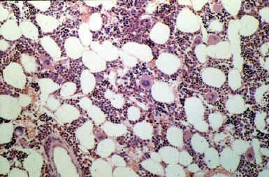
Histological section of porcine bone marrow at the stroma level (200X) in which various populations of hematopoietic cells can be observed.
|
THE PRIMARY LYMPHOID ORGANS consist of the bone marrow and the thymus. Their function is the production, regulation and differentiation of the different lymphocytes.
Maturing of the B lymphocytes takes place in the bone marrow and populations of T lymphocytes mature in the thymus.
Bone marrow: This is considered to be the main lymphopoietic organ in the pig, as well as in other mammals. It is distributed throughout the inside of all the bones of the animal, mainly in the long bones. The lymphopoietic stem cells are produced here, which will later give rise to macrophages and B and T lymphocytes. The latter T-cells migrate from the embryonic bone marrow to the cortex of the thymus.
From a structural and functional point of view, pig bone marrow is made up of two large interrelated compartments:
|
|
Bone marrow stroma is made up of a framework of fibres and reticular cells whose role is the support the hematopoietic part of the bone marrow, as well as the production of different growth and development factors to activate the precursor cells produced in the hematopoietic part. The stroma has been defined as the hematopoietic microenvironment necessary for the productive part of the bone marrow to function correctly.
The hematopoietic part, found in the parenchyma of the bone marrow, is formed by cell colonies or nests which are located in the networks of reticular fibres. The stem cells are known as "Colony Forming Units". These cells give rise to different cell lines, representing the main hematopoietic organ. It is still not well-known how the path is started towards cell differentiation to become one type of cell or another.
As well as the bone marrow, pig B lymphocytes are also produced in smaller quantities in other lymphoid organs associated with the mucosae, and in the spleen.
|
|
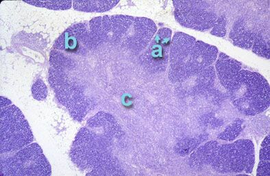
Histological section of pig thymus (25 X) in which a lobe can be observed with the different lobules separated by septae (a), the cortical zone or cortex (b) with a high presence of lymphocytes, the medullar zone or medulla (c) made up of epithelial cells and a small number of lymphocytes.
|
The thymus is a very important organ in the development of the immune system during the embryonic stage as it plays an important role in the selection of self lymphocytes (associated to the SLA of each animal), as well as during the first months of life of the animal. In a young pig it extends caudally from the digastric muscle, along the carotid arteries, to both sides of the neck, until the thoracic opening where both sides of the thymus join. This organ reaches its maximum size between the fifth and eighth month of the animal's life and gradually disappears after the first year of life.
In pigs, the thymus has a structure made up of different lobes surrounded by a thin layer of connective tissue, which continues into fine septae which subdivide the lobes into several partially separated lobules. Histologically and functionally they are differentiated into two zones: A cortex essentially constituted of a large number of proliferating lymphocytes of different sizes (between 9 and 5 microns) and a smaller number of epithelial cells, and a medulla made up of numerous epithelial cells which form concentric structures known as Hassall's corpuscles, and T lymphocytes in a lower number than in the cortex.
The thymus is the first lymphoid organ which develops from the stem cells of the bone marrow. Once in the thymus, these cells differentiate into T lymphocytes, remaining in the thymus or migrating to other secondary lymphoid organs, forming the T-dependent zones.
The learning process of the lymphocytes to acquire self-tolerance and the capacity to react against foreign molecules mainly takes place as they pass from the cortical zone to the medulla in the thymus. |
|
THE SECONDARY LYMPHOID ORGANS are formed:
|
|
|
|
| The main function of these organs is to recognize antigens and initiate the immune response. |
|
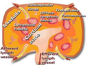
Diagram of pig lymph node in which it can be seen that its cell distribution and circulation is different to that of other mammals. The cortical zone with its lymphoid follicles and diffuse lymphoid tissue is found in the central area of the node; this is the medulla area in the periphery.
|
At systemic level:
Lymph nodes: The lymph nodes are nodular formations of dense connective tissue and whitish colour which are interspersed along the course of the lymphatic vessels forming the ganglion chains. Their function is to detain the antigens that may arrive from the lymphatic fluid and proceed to antigenic presentation and processing by collaboration with the macrophages and the lymphocytes of which the fluid is made.
The pig has a different lymphatic circulation and cellular disposition to that of other mammals. The lymph penetrates the concave surface (afferent lymphatic vessel) and leaves by the convex surface (efferent lymphatic vessel). |
|
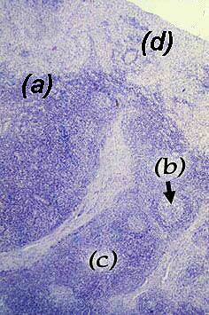
Histological section of pig thymus (25 X) in which the different distribution compared to other animal species can be observed; the cortical zone (a) is located in the centre of the node with a high presence of lymphoid follicles (b) (CD4+ B and T lymphocytes) and diffuse lymphoid tissue (c) and the medullary zone in the periphery (d)
|
Histologically and functionally-speaking, two well distinguished areas are observed in the lymph node parenchyma: cortical tissue and medullary tissue.
The cortical tissue is found in the central area of the nodular units and in the subcapsular areas, surrounding the efferent hilus. It consists of lymphoid follicles and diffuse lymphoid tissue. In turn, the lymphoid follicles consist of the germinal centre mainly comprising B lymphocytes and several populations of T lymphocytes (CD4+). It is here where the selection of high affinity B lymphocytes to react with the antigen takes place.
The diffuse lymphoid tissue is considered as a T dependent zone. It has a stroma made of fibres and reticular cells forming a three-dimensional network where, as well as T lymphocytes, macrophages and dendritic cells can be seen.
The medullary tissue is distributed by the periphery of the nodular units and around the efferent hilus. It mainly consists of fixed cell elements with reticular fibres and collagen. In contrast to other species it has neither medullary cords nor sinuses. It forms a uniform and diffuse framework with less permeability than other species. It is poor in lymphocytes, although macrophages and dendritic cells can be observed |
|
| The spleen is a secondary lymphoid organ, found on the left side of the abdominal cavity. It has an elongated and narrow shape and is bright red in colour. This lymphoid organ has a double function. Firstly, it works as a filter at blood level (the lymph nodes are filters at lymphatic level) for antigen recognition (immune function). Secondly, it serves as storage for blood cell production, mainly erythrocytes and platelets (hematopoietic function). |
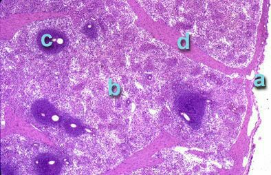
Histological section of pig spleen (100 X) in which the capsule (a), red pulp (b), white pulp (c) and trabeculae (d) can be observed.
|
The pig’s spleen has a framework made of a fine capsule of smooth muscle fibres, from which emerge trabeculae of smooth muscle fibre and collagen supporting the functional part of the spleen, consisting of: the splenic white pulp and the splenic red pulp.
The splenic white pulp is the lymphoid tissue of the spleen, always distributed around an artery or arteriole, forming lymphoid follicles and periarterial lymphoid sheaths. The lymphoid follicles are made up of: lymphocytes (mainly B), some plasma cells and antigen-presenting cells (macrophages, dendritic). The periarterial lymphoid sheaths are however made up of T lymphocytes, with some B lymphocytes being found in the more peripheral zones. One significant characteristic of the pig spleen is that within T lymphocyte populations, the CD4+ populations are much more numerous than CD8+. |
|
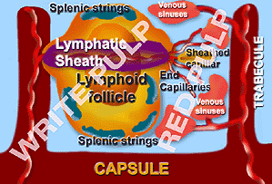
This diagram shows the different structural components of the spleen. The white pulp, formed by the lymphoid follicles and the periarterial lymphoid sheaths and the red pulp with its venous sinuses, splenic strings (or s. cords) and sheathed capillaries. All of this is protected by the capsule.
|
The splenic red pulp in the pig consists of three different structures: The splenic cords, the venous sinuses and the sheathed capillaries.
The splenic cords of the pig are made up of a framework of muscle fibres which support a large amount of free cells from the bloodstream such as monocytes, erythrocytes and platelets. The other cell population observed is that of fixed macrophages which are attached to the muscle fibres. The venous sinuses of the pig are wide and anastomotic ducts which lack fenestration in the basal membrane, allowing the splenic macrophages to perform the task of phagocytosis more easily. The sheathed capillaries are discontinuous capillaries which exhibit thickening of their walls, known as "ellipsoid" or "periarterial macrophage sheath" which are highly developed in pigs. They consist of many macrophages. |
|
| At mucosa level: Mucosal-associated lymphoid tissue. This is the immune system located throughout the mucosae, which is relatively independent of the systemic immune system. Throughout the pig’s intestine this is called gut associated lymphoid tissues (GALT).
Mucosal-associated lymphoid tissue is made of nodes of lymphoid tissue responsible for protecting the mucosae of the pig from attack by pathogens at local level. The immune system of the mucosae plays a very important role as in pigs many pathogens use the mucosae as entry pathways. About 80% of their action mechanisms are mediated by immunoglobulins of the IgA isotype, except in the tonsils where the synthesized immunoglobulin found in greatest quantities is IgG, followed by IgA. The most organized structures of GALT in pigs are:
|
|
|
The tonsils are part of the secondary lymphoid organs. Because of their location in the soft palate between the respiratory and digestive tract, they form part of the immune system of the respiratory and digestive mucosae.
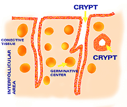
They are of key importance in the defensive mechanisms of the pig against infectious agents. They are made of a single node or group of nodes and diffuse lymphoid tissue. They are found in the lamina propria, closely linked to the epithelium, which according to its location is: stratified (oropharyngeal area) or pseudostratified in the nasopharyngeal area. Diagram of pig’s tonsils. The internal distribution formed by the crypts can be observed, with a different appearance according to the section, and the lymphoid follicles, with their germinal centres and the interfollicular tissue or diffuse tissue. |
|
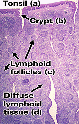
Histological section of pig’s tonsil (25 X) showing: stratified epithelium (a), crypts (b), lymphoid follicles (c) (mainly consisting of B lymphocytes) surrounded by diffuse lymphoid tissue (d) formed mainly of T lymphocytes.
|
The surface of the tonsils can be smooth or with deep invaginations, such as in the case of the epiglottic tonsil of the pig, allowing a large amount of lymphoid cells to concentrate in the invaginations.
The lymphoid structure of the tonsils is similar to that of the lymph nodes. They are made of lymphoid follicles with germinal centres, which are confined and compact groupings made of a large amount of B lymphocytes found within the diffuse lymphoid tissue which is mainly formed of T lymphocytes. All of this is closely linked to the epithelium.
Mainly IgG is produced in the tonsils, followed by IgA Peyer’s patches. These are groups of non-encapsulated lymphoid tissue, and located in the submucosa of the intestine. Two types can be distinguished in pigs by their size and location:
|
|
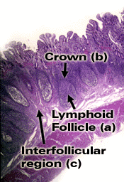
Histological section of Peyer’s patches (25 X). Here can be observed the lymphoid follicles with their germinal centre (a), the crown (b), the interfollicular region (c).
|
Histologically and functionally four areas can be distinguished in the Peyer’s patches:
- The lymphoid follicles.
- The crown.
- The interfollicular region.
- The dome.
- The lymphoid follicles: They are located in the submucosa of the intestine. They have great lymphopoietic activity, mainly in the germinal centres.
- The crown: This is the area where the lymphoid follicle and the capsule join, formed by T and B lymphocytes.
- The interfollicular region: This is formed by post-capillary venules which enhance the circulation of lymphocytes.
-
The dome: This is the area incorporated in the intestinal mucosa which covers the lymphoid follicles. It is formed of T lymphocytes, above all CD4+, B lymphocytes, macrophages and dendritic cells. At dome level, the intestinal mucosa is modified and villi and crypts disappear, the epithelium does not have caliciform cells and some M cells appear which have the role of antigenic presentation.
|
|
Jejunal Peyer’s patches: These are small patches (from 25 to 35) distributed throughout the jejunum and proximal section of the ileum and which persist throughout the life of the pig. They are made of B and T lymphocytes.
Ileocecal Peyer’s patches: These are large in size, and are located in the terminal section of the ileum. They develop during the first year of the animal’s life. Their cell composition also differs from that of the jejunal patches. In the ileocecal patches there are ten times more B lymphocytes than T lymphocytes. |
|
It is also important to point out that throughout the pig’s digestive tract, above all in the stomach and large intestine, at both mucosa and submucosa level, isolated lymphoid follicles can be observed. These are covered with an epithelium similar to that of the Peyer’s patches and whose function also appears to be similar. |
|